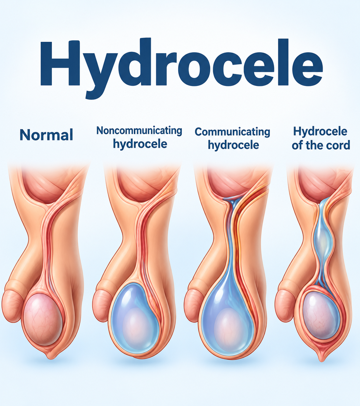
A hydrocele is a painless collection of fluid around the testicle that makes one side of the scrotum swell. In babies and young children, most hydroceles are present from birth and disappear on their own within the first one to two years, so watchful waiting is the usual first step. In adults, hydroceles are usually acquired later in life, rarely resolve without treatment, and require an ultrasound to rule out an underlying cause before any decision about surgery. The name is the same, but the cause, the timing of treatment, the operation itself, and the expected outcome differ substantially by age.
What is a hydrocele?
The testicle is wrapped in a thin, two-layered membrane called the tunica vaginalis — a sac that normally contains only a few drops of lubricating fluid. When fluid builds up between these two layers, the result is a hydrocele.
The typical sign is a smooth, painless swelling of the scrotum. Larger hydroceles can cause a dragging or heavy sensation, discomfort when walking, and difficulty with clothing. Pain is not a usual feature. Sudden pain, redness, fever, or a firm lump felt on the testicle itself point to a different problem and need prompt assessment.
Hydroceles are common at both ends of life. They are frequently noticed in newborn boys, and in adults they are seen most often in men over 40. Because the underlying mechanism differs between these two groups, it is useful to consider them separately.
Why hydroceles form: children vs adults
Children: a passage that has not fully closed
Before birth, the testicles develop inside the abdomen and then descend into the scrotum. As each testicle moves down, it pulls a finger-like extension of the abdominal lining with it. This channel is called the processus vaginalis, and it normally seals off around the time of birth.
If the channel stays open, fluid from the abdomen can flow down into the scrotum. This is called a communicating hydrocele. Its hallmark is a change in size during the day: the swelling is often small in the morning and larger in the evening after the child has been upright and active. If the opening is wide enough for a loop of bowel to pass through, the child has an inguinal hernia — a bulge in the groin — rather than fluid alone.
When the channel has closed but some fluid remains trapped around the testicle, the hydrocele is non-communicating. These do not change much in size and are especially likely to be absorbed over time.
Adults: an imbalance between fluid production and absorption
In adults, almost all hydroceles are non-communicating. The tunica vaginalis keeps producing a small amount of fluid while also absorbing it. When production exceeds absorption, fluid slowly accumulates over months or years.
Most adult hydroceles are idiopathic, meaning no specific cause is found. A smaller proportion are secondary hydroceles that develop alongside another condition. Recognized triggers include epididymitis (infection or inflammation of the coiled tube behind the testicle), scrotal injury, previous surgery for a varicocele, and, importantly, testicular cancer. Published case reports describe tumors that went unnoticed for some time because a large hydrocele hid the testicle from physical examination. This is the main reason an adult with a new hydrocele should have a scrotal ultrasound.

Key differences at a glance
The table below summarizes how a hydrocele in a child differs from one in an adult.
| Feature | Children | Adults |
| Main cause | Patent processus vaginalis (congenital) | Fluid production–absorption imbalance (acquired) |
| Common types | Communicating and non-communicating | Mostly non-communicating |
| Spontaneous resolution | Common, especially in infancy | Uncommon |
| Main condition to rule out | Inguinal hernia | Testicular tumor, epididymitis, spermatocele |
| Surgical approach | Small groin incision or laparoscopy | Incision in the scrotum |
| Goal of surgery | Close the open channel | Treat the sac so fluid cannot re-accumulate |
How is a hydrocele diagnosed?
Diagnosis starts with a physical examination. A clinician may shine a light through the scrotum, a test called transillumination. Clear fluid lets the light glow through, whereas a solid mass does not. Comparing the size of the swelling when lying down and when standing helps indicate whether the hydrocele communicates with the abdomen.
Scrotal ultrasound is the standard imaging test. It shows how much fluid is present, confirms that the testicle and epididymis look normal, and checks for a hernia. In adults, ultrasound is recommended for nearly every new hydrocele because the testicle cannot always be felt clearly through the fluid. In children, ultrasound is used when the nature of the swelling is uncertain or when an abdominoscrotal hydrocele is suspected. This uncommon variant extends from the scrotum up into the lower abdomen and can place pressure on the testicle.
Hydrocele in babies and children: when to wait and when to operate
The 2024 update of the European Association of Urology (EAU) and European Society for Paediatric Urology (ESPU) guidelines recommends observation as the initial approach for hydroceles in children. There is no convincing evidence that a simple hydrocele damages the testicle while it is being observed.
The likelihood of spontaneous resolution falls as the child gets older. A 2020 study in Pediatric Surgery International followed 113 Japanese children with asymptomatic hydroceles without immediate surgery. Resolution rates by age at first presentation were:
| Age at presentation | Spontaneous resolution |
| 1 month or younger | 94.3% |
| 1 to 12 months | 75.0% |
| 12 to 24 months | 65.0% |
| Older than 24 months | 33.3% |
In practical terms, nearly every hydrocele noticed in the first month of life went away without treatment. Even after age two, about one in three still resolved. In a subgroup of children, testicular size measured before and after the observation period did not change. This is a single retrospective study from one country, so the exact percentages should not be applied rigidly to every child, but the overall pattern matches other published series.
When surgery is usually considered in children
- A communicating hydrocele that persists beyond roughly 1 to 2 years of age
- An associated inguinal hernia
- A hydrocele that keeps enlarging or becomes tense
- An abdominoscrotal hydrocele that may compress the testicle
How the operation is performed in children
Because the problem in children is an open channel, surgery aims to tie off and divide the processus vaginalis at the level of the groin. This is done through a short incision in a natural skin crease of the lower abdomen or, in some centers, laparoscopically through tiny keyhole incisions. The operation is usually performed under general anesthesia as a day-case procedure. The adult-style operation, in which the scrotum is opened and the sac is excised, is not the standard approach for a communicating hydrocele in a child.
Hydrocele in adults: symptoms decide whether to treat
An adult hydrocele seldom disappears on its own, but that alone is not a reason for surgery. A hydrocele does not, by itself, impair testicular function. If the swelling is small and causes no discomfort, it can be monitored once ultrasound has excluded an underlying cause.
Treatment is considered when the hydrocele becomes large enough to cause discomfort, interfere with walking, exercise, or work, or create a cosmetic concern. A rapidly enlarging hydrocele, or one that appears after an infection or injury, also warrants closer evaluation.
Hydrocelectomy and other adult surgical options
The standard operation in adults is a hydrocelectomy — surgical treatment of the fluid-filled sac through an incision in the scrotum. Several techniques exist. They differ mainly in how the sac is handled after the fluid is drained.
| Technique | What is done | Practical notes |
| Sac excision | Most of the sac wall is removed and the edges are sealed | Suits large hydroceles and thick sac walls; bleeding control is important |
| Jaboulay (eversion) | The sac is turned inside out and stitched behind the testicle | Long-established standard technique |
| Lord (plication) | The sac is gathered with a series of stitches without excision | Less tissue dissection; suits thin-walled sacs |
| Minimal-incision techniques | A small window of the sac is treated through about a 2-cm cut | Can be performed under local anesthesia |
| Aspiration and sclerotherapy | Fluid is drained with a needle and a scarring agent is injected | Nonsurgical; higher recurrence than surgery |
What the evidence shows
There is no single best technique for every patient. The choice depends on the size of the hydrocele, the thickness of the sac, the patient’s bleeding risk, and the surgeon’s experience.
A 2019 study compared 276 operations using three open techniques. The overall recurrence rate was about 6%, with no significant difference between methods. The Lord technique had the lowest rates of overall complications and of hematoma, a collection of blood in the scrotum after surgery. A 2020 randomized trial in International Urology and Nephrology found that sac excision using a vessel-sealing device produced less postoperative swelling and higher patient satisfaction than the Jaboulay technique.
Newer data focus on less invasive surgery. A 2026 study in Translational Andrology and Urology reviewed 313 men treated by several surgeons in a U.S. multi-surgeon cohort. Complications occurred in 6.1% after a minimal-incision technique and in 13% after standard hydrocelectomy. Bleeding complications were seen in 6.1% of standard operations and in none of the minimal-incision cases. One-year recurrence was similar in both groups. For patients taking blood thinners, this difference in bleeding risk may be clinically meaningful. Longer follow-up is still needed to confirm that recurrence remains comparable over time.
Aspiration and sclerotherapy
Simple needle aspiration — drawing off the fluid without further treatment — is not recommended, because the fluid almost always returns. Aspiration followed by sclerotherapy adds an injected agent that causes the two layers of the sac to scar together. A Cochrane systematic review of randomized trials, involving 275 patients, found that sclerotherapy caused fewer complications, cost less, and allowed a faster return to work, but led to more recurrences than surgery. The review judged the overall evidence to be limited.
Some urologists use sclerotherapy cautiously in younger men who plan to father children. The sclerosing agent may affect the nearby epididymis, the structure that carries sperm away from the testicle.
Recovery and long-term outlook after hydrocele surgery
Children
Hydrocele repair in children has a high success rate, and recurrence is uncommon. Rare complications include injury to the vas deferens (the sperm duct) or to the blood supply of the testicle. For this reason, the operation is best performed by a surgeon experienced in pediatric groin surgery. Most children go home on the day of surgery.
Adults
The most common issues after adult surgery are scrotal swelling and hematoma. Swelling settles gradually over several weeks. During this time, supportive underwear and cold packs help with comfort. Infection, persistent scrotal pain, and recurrence are less frequent but possible.
The epididymis lies close to the sac, and injury during surgery could affect the passage of sperm. Men who plan to have children should discuss this with their surgeon before choosing a technique. If a hydrocele comes back, it can be treated with repeat surgery or, in selected cases, sclerotherapy.
When to see a doctor
Talk to your doctor, who may refer you to a urologist or pediatric surgeon, if any of the following apply:
- A baby’s scrotal swelling persists beyond 1 to 2 years of age
- A bulge appears in the groin, especially one that comes and goes
- Scrotal swelling is accompanied by pain, redness, or fever
- An adult develops new scrotal swelling, or an existing swelling grows quickly
- A firm lump can be felt on the testicle itself
Sudden, severe scrotal pain can indicate testicular torsion, a twisting of the testicle that cuts off its blood supply. This is a surgical emergency that requires immediate care.
Frequently asked questions
Can a hydrocele go away on its own?
In infants, yes, and commonly so. Most hydroceles found in the first months of life resolve without treatment, and some still resolve after age two. In adults, spontaneous resolution is uncommon, so the hydrocele is monitored for changes in size and symptoms.
Does a hydrocele cause infertility?
There is little evidence that a hydrocele itself reduces sperm production. The more relevant concern is the small risk of injuring the epididymis during surgery or sclerotherapy. Men planning a family should raise this with their urologist when choosing a treatment.
Why not just drain the fluid with a needle?
Needle drainage alone is followed by recurrence in most cases, because the sac keeps producing fluid. Combining aspiration with sclerotherapy improves results, but recurrence remains more frequent than after surgery.
How long does recovery take after adult hydrocele surgery?
Most adults resume light daily activities within a few days. Swelling can take several weeks to settle completely. Strenuous exercise and cycling are usually postponed until the swelling has gone down. The exact timing depends on the size of the hydrocele and the technique used.
Is hydrocele surgery in children done through the scrotum?
Usually not. A child’s hydrocele is typically caused by an open channel from the abdomen, so the repair is performed through a small groin incision or laparoscopically. The goal is to close that channel rather than remove the sac.
Key takeaways
- Hydroceles in children are mostly congenital, and observation is the first step. Persistent hydroceles beyond 1 to 2 years, or those with a hernia, are repaired through the groin.
- Hydroceles in adults are mostly acquired. Ultrasound should exclude a tumor or infection, and surgery is reserved for symptomatic cases.
- Several adult techniques give similar recurrence rates of around 6%. Minimal-incision and plication methods appear to reduce bleeding-related complications.
- The overall outlook after treatment is good. Adults should expect several weeks of swelling and understand the small risk of recurrence.
If scrotal swelling persists or grows, talk to your doctor about whether a urology assessment is needed.
References
- Gnech M, van Uitert A, Kennedy U, et al. European Association of Urology/European Society for Paediatric Urology guidelines on paediatric urology: summary of the 2024 updates. Eur Urol 2024;86:447-56.
- Kurobe M, Harada A, Sugihara T, et al. The outcomes of conservative management and the natural history of asymptomatic hydroceles in children. Pediatr Surg Int 2020;36:1189-95.
- Shi C, Wei N, Schrup S, et al. Comparative outcomes of minimal-incision versus standard hydrocelectomy: reduced bleeding and healthcare utilization in a multi-surgeon cohort. Transl Androl Urol 2026;15:247.
- Özkaya F, Cakici OU. Jaboulay’s technique contrasted with a novel hydrocelectomy technique using a vessel sealer in the treatment of adult hydrocele: a prospective randomized study. Int Urol Nephrol 2020;52:447-53.
- Shakiba B, Heidari K, Jamali A, Afshar K. Aspiration and sclerotherapy versus hydrocoelectomy for treating hydrocoeles. Cochrane Database Syst Rev 2014;(11):CD009735.
This article is intended as general medical information and does not replace individual diagnosis or treatment. If you have symptoms, please consult a qualified healthcare professional.
Read together
Leave a Reply